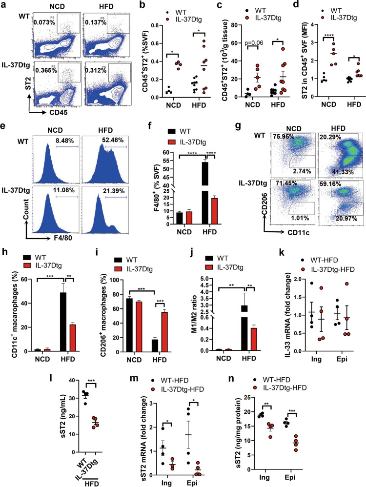
Fig. 4. IL-37D transgene amplifies ST2+ immune cell population and promotes M2 macrophage polarization in WAT via sST2 suppression in HFD-fed mice.
IL-37Dtg and WT mice were fed on HFD or NCD for 18 weeks (n = 4–8 per group). a–d SVF from inguinal WAT was analyzed by flow cytometry. Representative (a) and statistical data on the percentages of CD45+ST2+ cells (b), the numbers of CD45+ST2+ cells (c), and the mean fluorescence intensity (MFI) of ST2 (d) are shown. e–j SVF from epidydimal WAT was analyzed by flow cytometry. Representative (e) and statistical (f) data on the percentages of F4/80+ macrophages are shown. Representative (g) and statistical (h, i) data on the percentages of CD11c+ M1 macrophages and CD260+ M2 macrophages in F4/80+ cells are shown. The ratio of M1 to M2 (j) was calculated. k–n The mRNA levels of IL-33 in WAT were determined by qPCR (k). The serum level of sST2 (l) was measured by ELISA. The mRNA (m) and secretion (n) levels of sST2 in WAT explants were determined by qPCR and ELISA. Data represent mean ± SEM. *p < 0.05, **p < 0.01, ***p < 0.001, ****p < 0.0001 determined by two-way ANOVA (b, c, d, f, h–j) or student’s t test (k–n).

