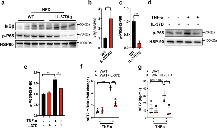
Fig. 6. Either endogenous or exogenous IL-37D blocks NF-κB activation and sST2 production in WAT.
IL-37Dtg and WT mice were fed on HFD for 23 weeks (n = 4 per group). The protein levels of IκB and p-P65 in inguinal WAT were detected by western blot (a) and the grayscale values are shown (b, c). d–g Epidydimal WAT explants from NCD-fed WT mice were treated with human IL-37D protein (10 ng/mL) in the presence or absence of TNF-α stimulation (20 ng/mL). The protein level of p-P65 was detected by western blot (d) and the grayscale values are shown (e). The mRNA (f) and secretion (g) levels of sST2 were detected by qPCR and ELISA. Data represent mean ± SEM. *p < 0.05, **p < 0.01, ***p < 0.001 determined by student’s t test (b, c), one-way ANOVA (e) or two-way ANOVA (f, g).

