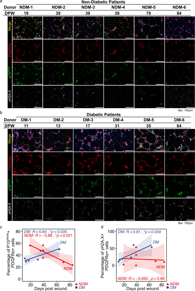
Fig. 5. Cellular senescence in subcutaneous adipose tissue during wound healing in diabetic patients.
a, b Representative images of adipose tissue immunostained for PDGFRα, p15INK4B, and γH2A.X. during wound healing in diabetic and non-diabetic patients. c, d Correlation between the percentage of p15INK4B + or γH2A.X + cells in PDGFRα + cells and time post-wounding (n = 6 for each group). Correlations were examined statistically using Pearson’s correlation coefficient, and 95% confidence intervals are shown with translucent fill corresponding to each marker color.

