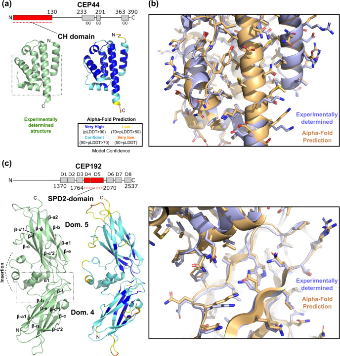
Fig. 1. AF2 predicts the structures of globular domains of centriolar proteins with high accuracy.
a Ribbon representation of the experimentally determined high-resolution structure of the N-terminal CH domain of CEP44 (PDB 7PT5) and the structure of CEP44’s CH domain as predicted by AF2. The AF2 prediction is colour-coded by the pLDDT values indicating the confidence level of the prediction. The dotted box indicates the region shown magnified in panel b. In the domain organisation scheme of human CEP44, cc denotes a predicted coiled coil and amino acid residue numbers indicate domain boundaries. b Detailed view of the region boxed in panel a, with the experimentally determined structure (blue) superposed with the AF2 structure prediction (orange). Sidechains are shown as sticks. c Comparison of the experimentally determined (PDB 7PTB) and the AF2-predicted structure of the so-called Spd2 domain of human CEP192, presented similarly as in panels a-b. D in the domain overview scheme indicates PapD-like domains. The beta strands of the two subdomains of the Spd2 domain are labelled according to the strand nomenclature of Bullock and colleagues61.

