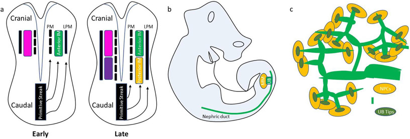
Figure 1. Diagrams of mammalian kidney development.
(a) A cranial-caudal axis in the primitive streak determines the laterality of gastrulating mesoderm, PM = Paraxial Mesoderm, LP = Lateral Plate Mesoderm. Early primitive streak forms anterior mesoderm while the late differentiates into posterior mesoderm.
(b) The ureteric bud (UB) originates from the anterior intermediate mesoderm (aIM) and develops as a caudal outpouching of the nephric duct to invade adjacent metanephric mesenchyme (MM), which is derived from the posterior intermediate mesoderm (pIM).
(c) Branching UB stalks terminate in distinct clusters of UB tip cells which invade nephron progenitor cells (NPCs) of the cap mesenchyme to initiate reciprocal induction.

