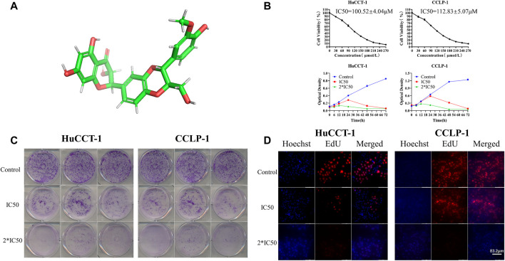
FIGURE 1.
Silibinin inhibits cell viability in cholangiocarcinoma cells. (A). Diagram of the three-dimensional structure of silibinin. (B). Silibinin inhibited cell viability of HuCCT-1 and CCLP-1 cell lines in a concentration-dependent and time-dependent manner. Cells were treated with varying concentrations of silibinin for indicated time points (6, 12, 24, 48 and 72 h) and cell viability was examined by the CCK-8 assay. (C). Colony formation assay results of HuCCT-1 and CCLP-1 cell lines treated with different concentrations of silibinin. (D). EdU proliferation assay results of HuCCT-1 and CCLP-1 cell lines treated with different concentrations of silibinin.

