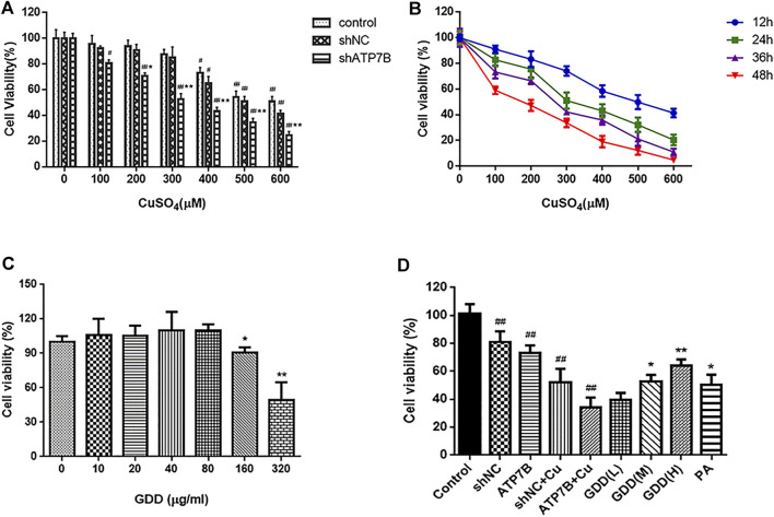
FIGURE 1.
(A) Cell viability in control, shNC, and shATP7B transfected after 24-h incubation with different concentrations CuSO4 ( ). # p < 0.05, ## p < 0.01 compared with corresponding blank group without CuSO4 treatment; * p < 0.05, ** p < 0.01 compared between shNC and shATP7B groups at same copper concentration; (B) dose and time-effect curves of shATP7B transfected cell viability after incubation with CuSO4 ( ); (C) cell viability after 24 h treated by GDD ranging from 0 to 320 μg/ml (0, 10, 20, 40, 80, 160, and 320 μg/ml) ( ). * p < 0.05, ** p < 0.01 compared with control group; (D) cell viability in copper-laden HLD hepatocytes before and after GDD treatment ( ). * p < 0.05, ** p < 0.01 compared with shATP7B + Cu group.

