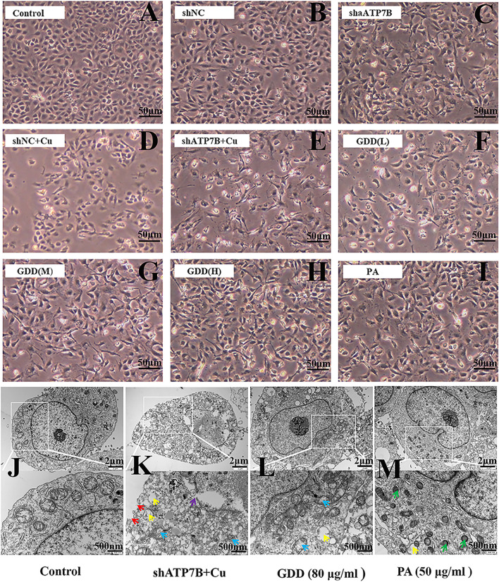
FIGURE 3.
(A–I) Morphological changes of copper-laden HLD hepatocytes treated with GDD by inverted microscope (×200); (J–M) ultrastructure of copper-laden HLD hepatocytes observed by transmission electron microscope (×10,000 or ×25,000) (red arrow: cell membrane rupture; yellow arrow: mitochondrial vacuolation; purple arrow: nuclear membrane rupture; blue arrow: mitochondrial cristal rupture; green arrow: mitochondrial shrinkage).

