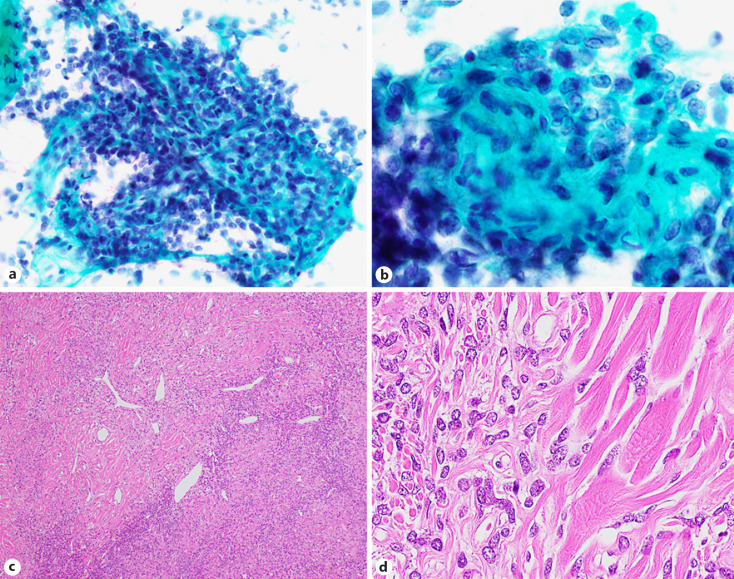
Fig. 4.
Cytological and histological features of an SFT (case 15). High-magnification images (a, b) show a cellular area composed of oval to short spindle-shaped cells with scant cytoplasm (×400). Hematoxylin-eosin staining (c) showed a patternless architecture with alternating hypo- and hypercellular areas and a prominent branching vasculature (×100). The higher magnification image of the hematoxylin-eosin staining (d) shows differently sized cuboidal to spindle-shaped cells within dense collagen (×400). SFT, solitary fibrous tumor.

