FIGURE 3.
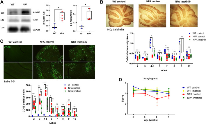
Acute imatinib treatment decreases neuronal death in the cerebellum and improves locomotor function in NPA mice. (A) WT and NPA brain homogenates (50 μg protein/lane) from mice at 4 weeks old were analyzed by Western blot. The graph shows quantifications of p-c-Abl levels normalized by GAPDH and c-Abl levels. The number of animals was: WT = 3; NPA = 4; Student’s t-test: *p < 0.05. (B) WT and NPA mice were i.p. injected with imatinib (12.5 mg/kg) or vehicle from 3 weeks of age until 7 weeks of age. The Purkinje neuron marker calbindin was analyzed by immunohistochemistry. A quantification of calbindin-immunoreactive Purkinje cell bodies in cerebellar sections is shown. (C) CD68 marker was evaluated by immunofluorescence analysis in the cerebellum from WT and NPA mice. For (B,C), the number of animals was WT control = 3; NPA control = 5; NPA imatinib = 4. Images were taken with × 4 objective. ANOVA, Tukey post-hoc: *p < 0.05; **p < 0.01; ***p < 0.001; ****p < 0.0001 (D) Mice were treated for 4 weeks and motor coordination was assessed weekly by the hanging test. Data are shown as mean ± SEM. ANOVA, Tukey post-hoc: *p < 0.05, NPA control is statistically different from WT control and NPA with imatinib. The following number of animals was used: WT control = 11; WT imatinib = 10; NPA control = 8; NPA imatinib = 9. In the box-and-whisker plots, the center line denotes the median value, edges are upper and lower quartiles, whiskers show minimum and maximum values and points are individual experiments.

