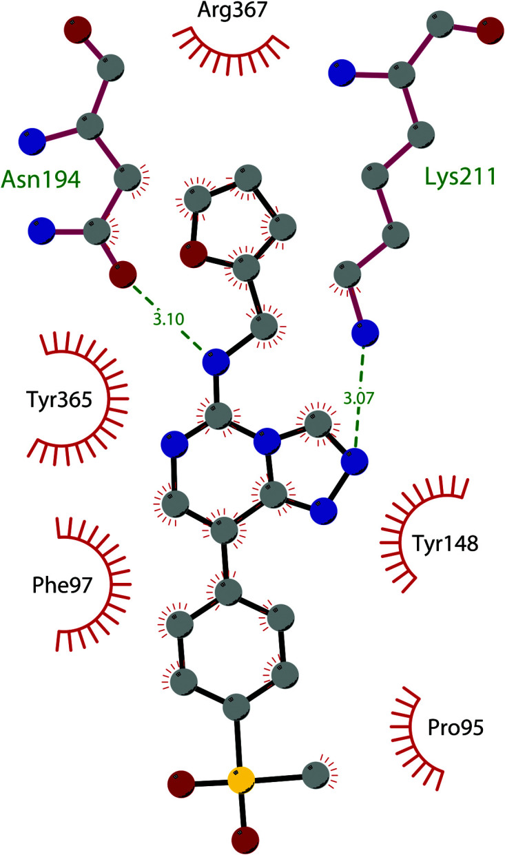
Fig. 14. Flat representation (generated using LigPlot+ from PDB ID 5GSA) showing contacts between compound 52, EED, and an EZH2 peptide referred to as the EED binding domain (EBD). Hydrogen bonds are shown as dashed lines. The spoked arcs represent protein residues making non-bonded (hydrophobic) contacts with compound.

