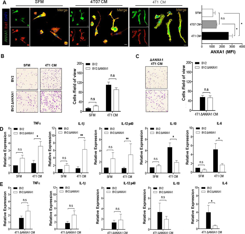
Fig. 5.
Extracellular and intracellular ANXA1 elicited different effects on migratory profiles in BV-2 microglial cells. a Primary adult microglia treated with either SFM, 4T07 CM, or 4T1 CM for 24 h and stained with ANXA1 (green), microglial marker Iba1 (red), and DNA-binding dye DAPI (blue). Graph (rightmost) showing the immunoreactivity against ANXA1 on primary adult microglial cells. b and c BV-2 or BV-2 ΔANXA1 microglia were treated with SFM, 4T1 CM or 4T1 ΔANXA1 CM for 24 h in the transwell migration assay. Representative brightfield images and quantification of migrated BV-2 or BV-2 ΔANXA1 microglia. c BV-2 or BV-2 ΔANXA1 microglia were treated with SFM, 4T1 CM or 4T1 ΔANXA1 CM for 24 h and gene expression of inflammatory markers were analyzed using qRT-PCR. Data represent mean ± SEM; n = 3 independent experiments. *P < 0.05, **P < 0.01

