Fig. 8.
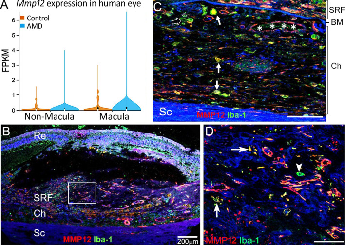
The expression of MMP-12 in human AMD and macular fibrosis. A The mRNA expression levels of Mmp12 in non-macular and macular RPE-choroid from non-AMD controls and AMD (Bulk RNA-Seq data from NCBI GEO database, ID: GSE135092). Mean ± SD, (AMD macula, n = 13; AMD non-macula, n = 10; non-AMD macula, n = 33; non-AMD non-macula, n = 36). B A confocal image from human nAMD with macular fibrosis showing MMP12 (red) and Iba-1 (green) in the choroid and subretinal lesion. C High magnification image showing Iba-1+MMP12+ cells (arrows), Iba-1+MMP12− cells (open arrow) in the choroid and MMP12+ cells in choroidal blood vessels (asterisks). D Magnified area of box in (B) showing Iba-1+MMP12+cells (arrows), Iba-1+MMP12−cells (arrowhead) in the macular fibrotic lesion. Re = retina, SRF = subretinal fibrotic lesion, Ch = choroid, BM = Bruch’s membrane. Scale bars in C and D = 50 µm

