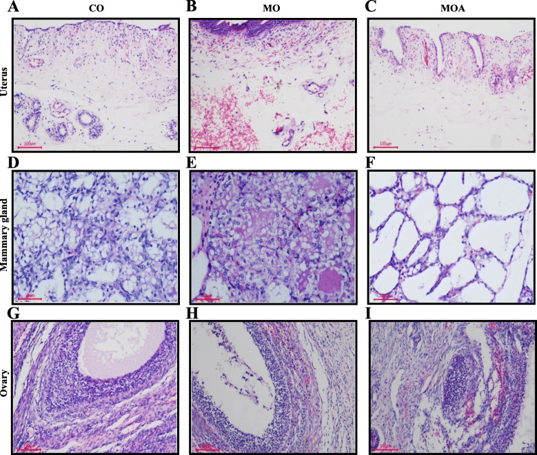
Fig. 1.
Effects of MBA on histopathology in organs of first-parity gilts when exposed to ZEA. A Uterus (CO group): a small amount of inflammatory cell infiltration in inner membrance; capillaries are mildly hyperemic or stagnant. B Uterus (MO group): a large amount of inflammatory cell infiltration in inner membrance; severe bleeding in lamina propria. C Uterus (MOA group): a small amount of inflammatory cell infiltration in inner membrance; capillaries are mildly hyperemic or stagnant. D Mammary gland (CO group): a small amount of reticular secretion in the acinar cavity; glandular epithelial cells swell and become fatty; occasionally, glandular epithelial cells exfoliate and become necrotic. E Mammary gland (MO group): a small amount of reticular or mass secretion in the acinar cavity; glandular epithelial cells swell and become fatty; A large number of glandular epithelial cells disintegrate from myoepithelial cell; the glandular cavity contains exfoliated and necrotic glandular epithelial cells. F Mammary gland (MOA group): no secretions found in the acinar cavity; glandular epithelial cells swell slightly; a small amount of steatosis occurs. G Ovary (CO group): normal structure; a small amount of eosinophils; no obvious histopathological changes observed. H Ovary (MO group): bleeding seen in the follicular membrane; a small amount of eosinophils; necrosis in the follicular granulocytes. I Ovary (MOA group): bleeding seen in the follicular membrane; a small amount of eosinophils

