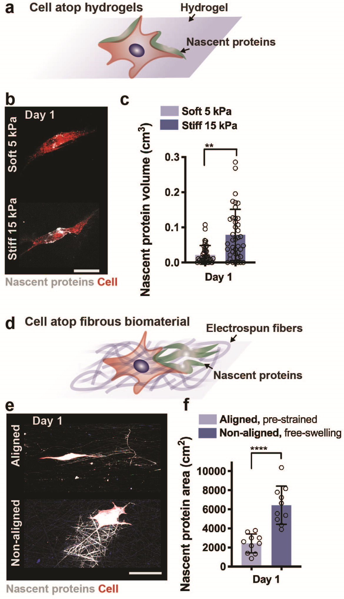
Figure 3. Application of nascent protein labelling to assess cell mechanobiologic response atop 2D biomaterials.

a Schematic illustrating the culture of a cell atop a synthetic hydrogel and accumulation of nascent proteins adjacent to the cell body. b Representative images (scale bar 50 μm) of nascent protein (AHA, grey) deposition by human mesenchymal stromal cells (hMSCs, cell membrane marker, red) seeded on non-degradable soft (5 kPa Young’s modulus) and stiff (15 kPa Young’s modulus) hyaluronic acid hydrogels and cultured for 1 day (24 hours) in growth media supplemented with the methionine-analog AHA. c Quantification of nascent protein volume deposited by hMSCs within 1 day of culture (n = 25 cells, mean ± SD, **p ≤ 0.01 by two-tailed Student’s t-test). d Schematic illustrating the culture of a cell atop a fibrous biomaterial and deposition of nascent proteins. e Representative images (scale bar 50 μm) of nascent protein (AHA, grey) deposition by bovine mesenchymal stromal cells (bMSCs, cell membrane marker, red) seeded on electrospun fibronectin-coated polycaprolactone fibers (aligned, pre-strained or non-aligned, free-swelling) and cultured for 1 day (24 hours) in growth media supplemented with the methionine-analog AHA. f Quantification of nascent protein area deposited by bMSCs within 1 day of culture (n = 10 cells, mean ± SD, ****p ≤ 0.0001 by two-tailed Student’s t-test). e,f adapted with permission from Bonnevie et al.21, Nature Research).
