
Figure 7. Quantification of nascent matrix thickness in ImageJ.
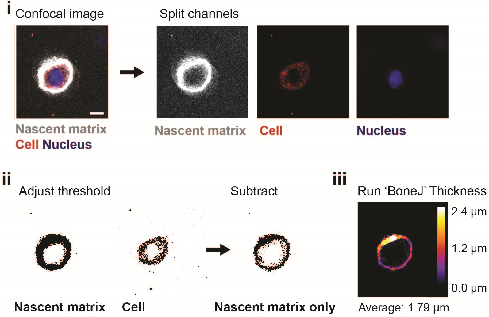
Acquire a confocal image of the nascent matrix, cell membrane and nucleus, and split the channels into single images using ImageJ (scale bar 10 μm). (i). To obtain an image of the nascent matrix only, adjust the threshold for each channel with ‘Otsu thresholding’ and subtract the ‘cell’ image from the ‘nascent matrix’ image (ii). Use the ImageJ plugin ‘BoneJ’ to measure the average nascent matrix thickness (iii).