. 2022 Mar 16;13(14):4150–4169. doi: 10.1039/d2sc00841f

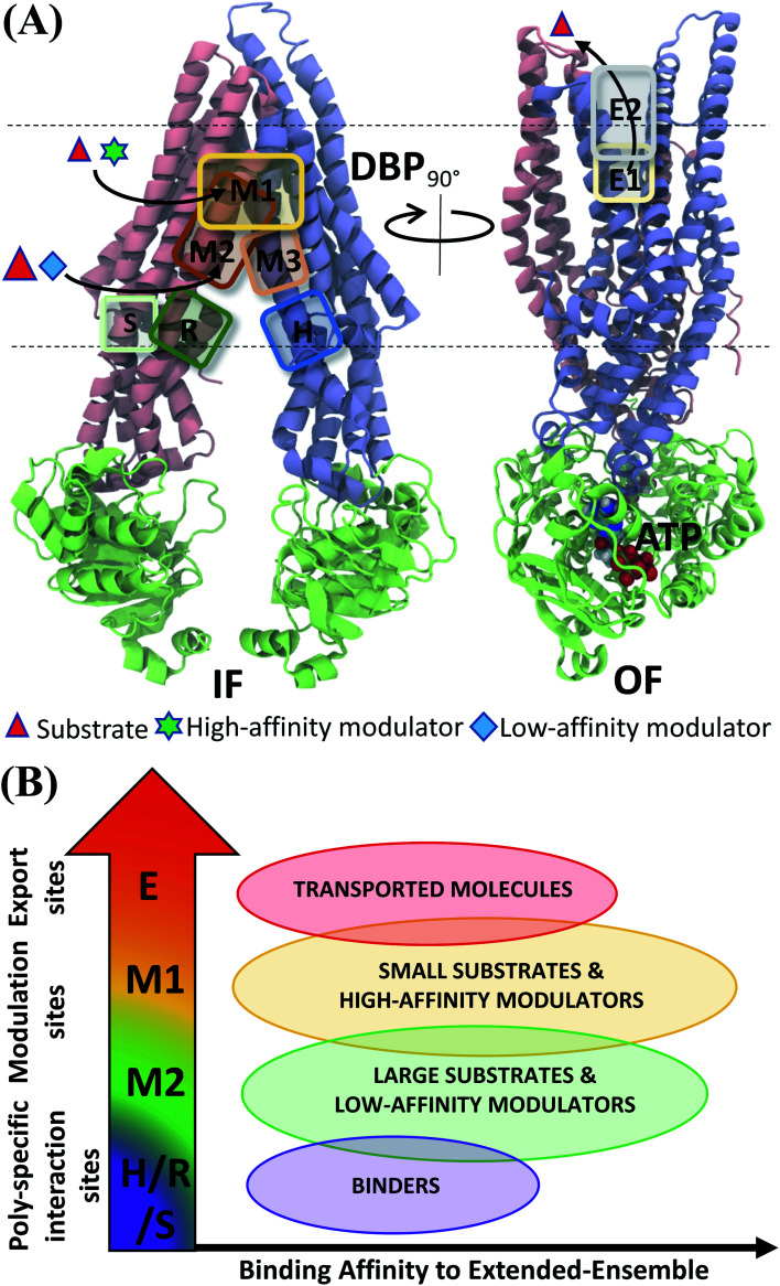
Fig. 11. Classification of ligand binding sites in Pgp identified by extended ensemble docking. (A) Different binding sites are shown in representative IF (left) and OF (right) conformations. The two TMD leaflets are shown in blue (TMD1) and pink (TMD2), respectively, and the NBDs are shown in green. The major substrate-modulator binding site (M1), as well as the low-affinity modulator/large substrate binding site (M2) are observed throughout the conformational transition of Pgp, whereas extracellular sites (E1 and E2) are only observed in the OF-like states. (B) Combining the predicted binding affinities of all compounds in the different binding sites obtained in the extended ensemble (Fig. 7 and S13), we observe differences in the relative binding affinities of the poly-specific interaction sites (H/R/S), modulation sites (M1/M2) and extracellular sites (E). These differences may facilitate the transport of the molecules from the inside to the outside of the cell.

Fig. 11