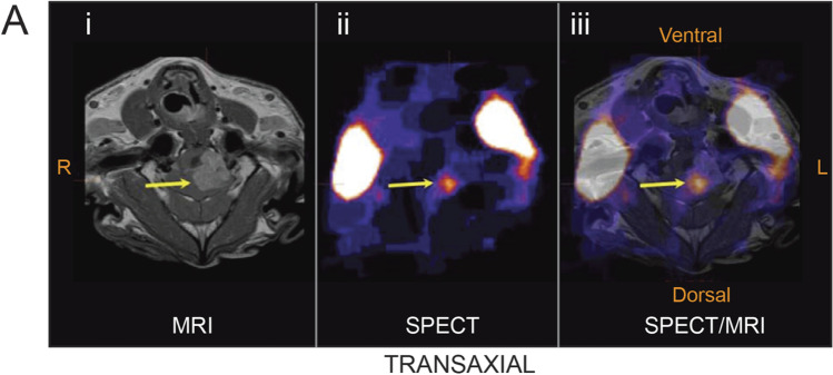
Fig. 5.
Biodistribution of systemically administered 123I labelled minicells Dox in dogs with brain cancer. Arrow showed the tumour location at 3 h post minicells administration. Minicell accumulation was evident with single-photon emission computed tomography (SPECT) imaging. Merge of MRI and SPECT illustrate the location of minicells at the core of brain tumour. Figure adopted with permission from MacDiarmid et al. (79).

