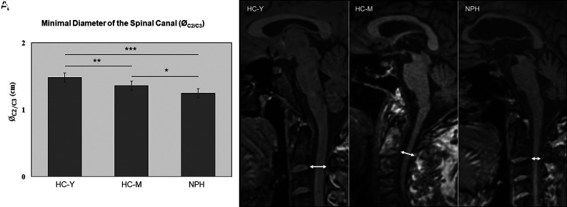
FIG 3.
Minimal diameter of the spinal canal (ØC2/C3). A, Minimal diameter of the spinal canal (ØC2/C3) in centimeters. B, Representative MR images (midsagittal T1-weighted image) to measure the minimal diameter of the spinal canal at the level of the intervertebral space between the second and third upper cervical vertebrae (double-sided arrow). The left panel shows an image of an HC-Y, the middle panel shows an HC-M, and the right panel shows an image of a patient with NPH. The asterisk indicates P < .05; double asterisks, P < .01; triple asterisks, P < .001.

