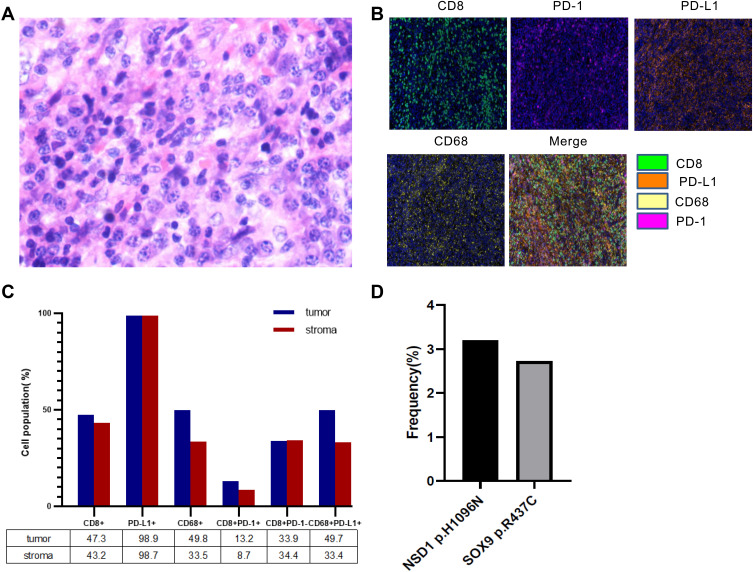
Figure 2.
(A) Hematoxylin and eosin staining of the recurrent lesions. (B) The expression of CD8, PD-1, PD-L1, and CD68 in resected tumor tissues was detected by multiple immunohistochemistry (mIHC). Nuclei (blue) were counter-stained by DAPI. Magnification ×200. (C) Quantification analysis of data in B. (D) The frequencies of two shared pathogenic mutations in the recurrent lesions.

