Fig. 3.
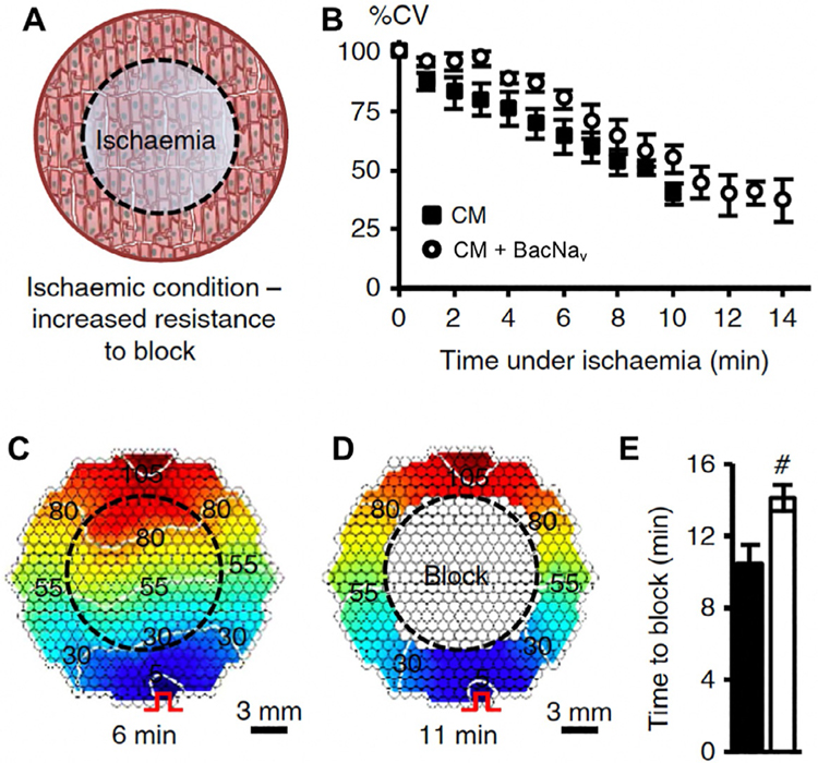
Improvement of mammalian AP conduction by BacNav in modeled pathological conditions. (A) Schematic depicting exogenous expression of BacNav in neonatal rat cardiomyocytes (CMs) to increase resistance to conduction block in ischemic conditions. Black dashed circle denotes position of glass coverslip used to induce regional ischemia in CM monolayer. (B) Progressive conduction slowing until block with time of ischemia in control (CM) and NavSheP D60A transduced monolayers. (C and D) Representative isochrone maps showing conduction slowing after 6min (C) and complete block after 11 min of ischemia (D). Pulse signs indicate location of stimulating electrode. (E) Under ischemic condition, CMs transduced with NavSheP D60A lentivirus (white) resisted conduction block longer than control CMs (black). Error bars indicate s.e.m.; statistical significance was determined by an unpaired Student’s t-test.

