
Figure 1.
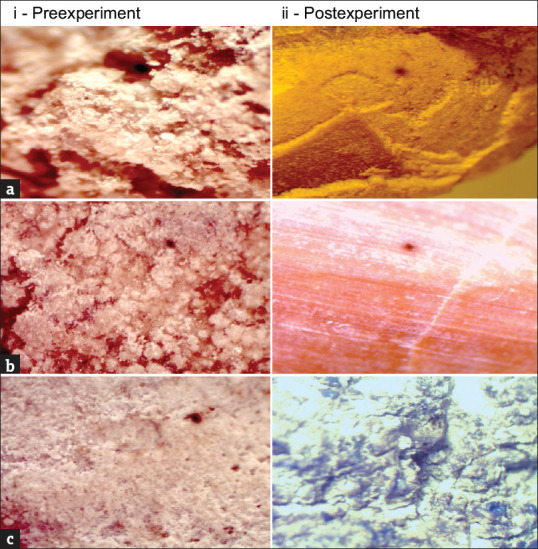
High-power microscopic pictures of calcium oxalate stone, uric acid stone, and combination of calcium oxalate and uric acid stone. (a) Calcium oxalate stone pictures taken pre- and post-experiments; pre-experiment picture shows hard stone brown in color which grow in radial fashion from the nidus with rounding wedge at extremity, and post-experiment picture shows disappearance of irregular surface probably due to dissolution exposing the interior table layer in a well-arranged pattern. (b) Uric acid stone pictures taken pre- and post-experiments; preexperiment picture shows dark orange stone composes of small spherical region, and postexperiment picture shows stone with smooth interior surface. (c) Calcium oxalate and uric acid stone pictures taken pre- and post-experiments; pre-experiment picture shows yellow cluster of platelets, and post-experiment picture shows nodular surfaces and discoloration of the stone