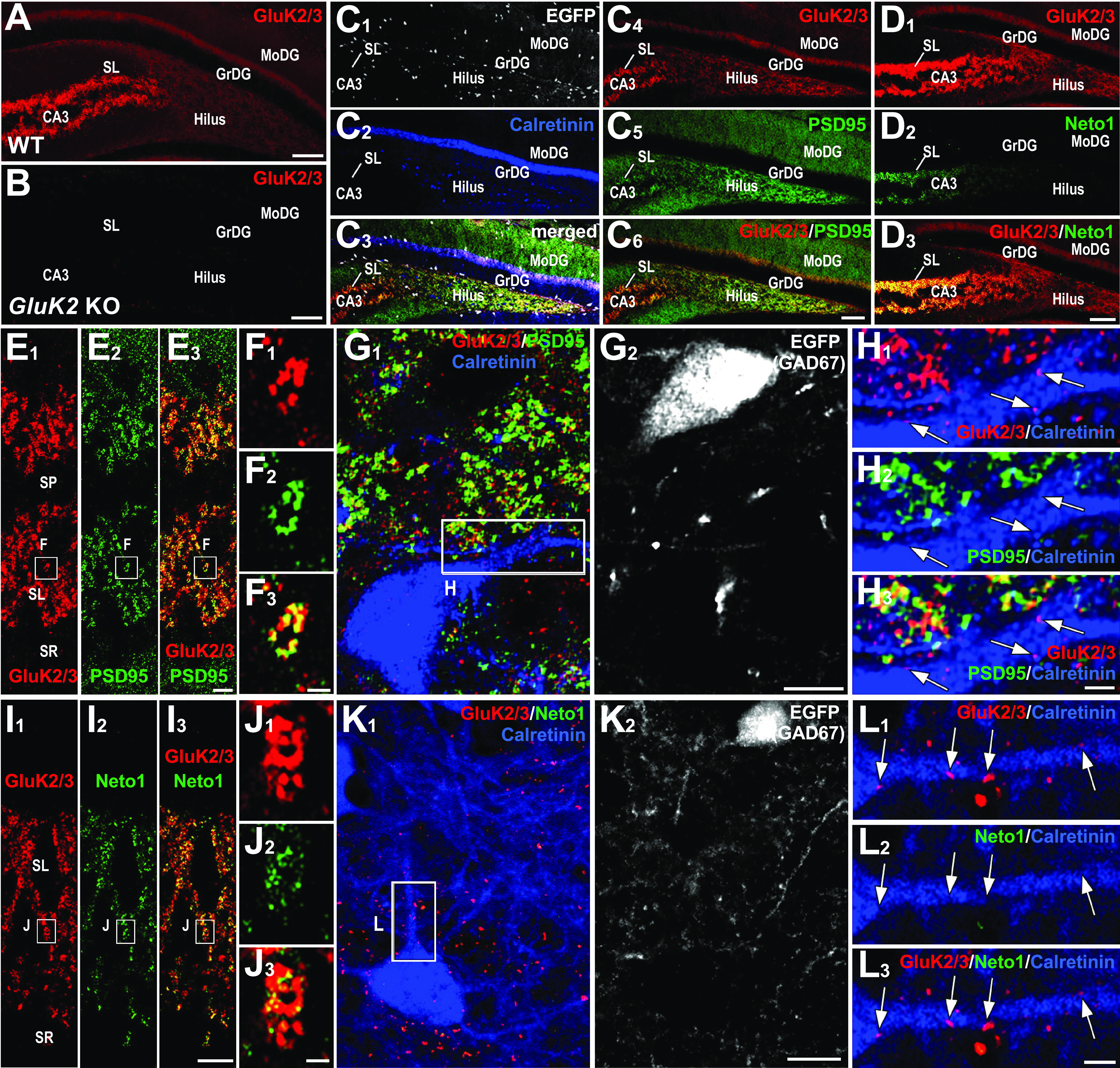
Figure 4.

Contrasting localization of GluK2/3 and its molecular partners in CA3 stratum lucidum and DG hilus. A, B, In WT mice, GluK2/3 labeling is intense in CA3 stratum lucidum, while it is moderate and diffuse in the dentate gyrus (A); note the lack of GluK2/3 staining in GluK2 KO mice, indicating the specificity of the GluK2/3 antibody and exclusive expression of GluK2 in these hippocampal regions (B). C1–C6, Quadplex immunofluorescence for GFP (C1, C3, white), calretinin (C2, C3, blue), GluK2/3 (C3, C4, C6, red), and PSD-95 (C5, C6, green) in GAD67+/GFP mice. D1, D2, Double immunofluorescence for GluK2/3 (D1, red) and Neto1 (D2, green). Note that intense signal for Neto1 is almost limited to CA3 stratum lucidum. E1–H3, Distinct GluK2/3 and PSD-95 localization between CA3 pyramidal cells and hilar MCs. E1–F3, GluK2/3 and PSD-95 labeling are intense in the CA3 stratum lucidum (E1–E3), and a high-magnification image confirms their extensive overlap (F1–F3). G1–H3, An MC, which is identified as a calretinin-positive (G1, blue) and GFP-negative (G2, white) cell in GAD67+/GFP mice, shows weak labeling for GluK2/3 on its dendrites (red, arrows in H1–H3). Note that such GluK2/3 puncta are neither overlapped nor associated with PSD-95 signal (H2, green). I1–L3, Distinct GluK2/3 and Neto1 localization between CA3 pyramidal cells and hilar MCs. I1–I3, Intense GluK2/3 and Neto1 labeling in the CA3 stratum lucidum (l). J1–J3, A high-magnification image shows that Neto1 labeling is observed only on GluK2/3-positive puncta. K1–L3, An MC, which is calretinin-positive(K1, blue) GFP-negative (L2, white) cell inGAD67+/GFP mice, shows weak labeling for GluK2/3 on its dendrites (K1, L2, L3, red arrows) but lacks Neto1 labeling (K1, L2, L3, green). GrDG, GC layer of the DG; MoDG, molecular layer of the DG; SL, stratum lucidum; SP, stratum pyramidale; SR, stratum radiatum. Scale bars: A, B, C1–C6, D1–D3, 100 µm; E1–G2, I1–K2, 10 µm; H1–H3, L1–L3, 2 µm.
