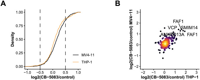
Fig 2. Dynamics of the ubiquitin-modified proteome in AML cells after VCP inhibition.
(A) SILAC ratio distribution of all quantified ubiquitylation sites in MV4-11 and THP-1 cells. (B) Scatter plot showing SILAC ratios of quantified ubiquitylation sites in MV4-11 and THP-1 cells after treatment with the VCP inhibitor CB-5083.

