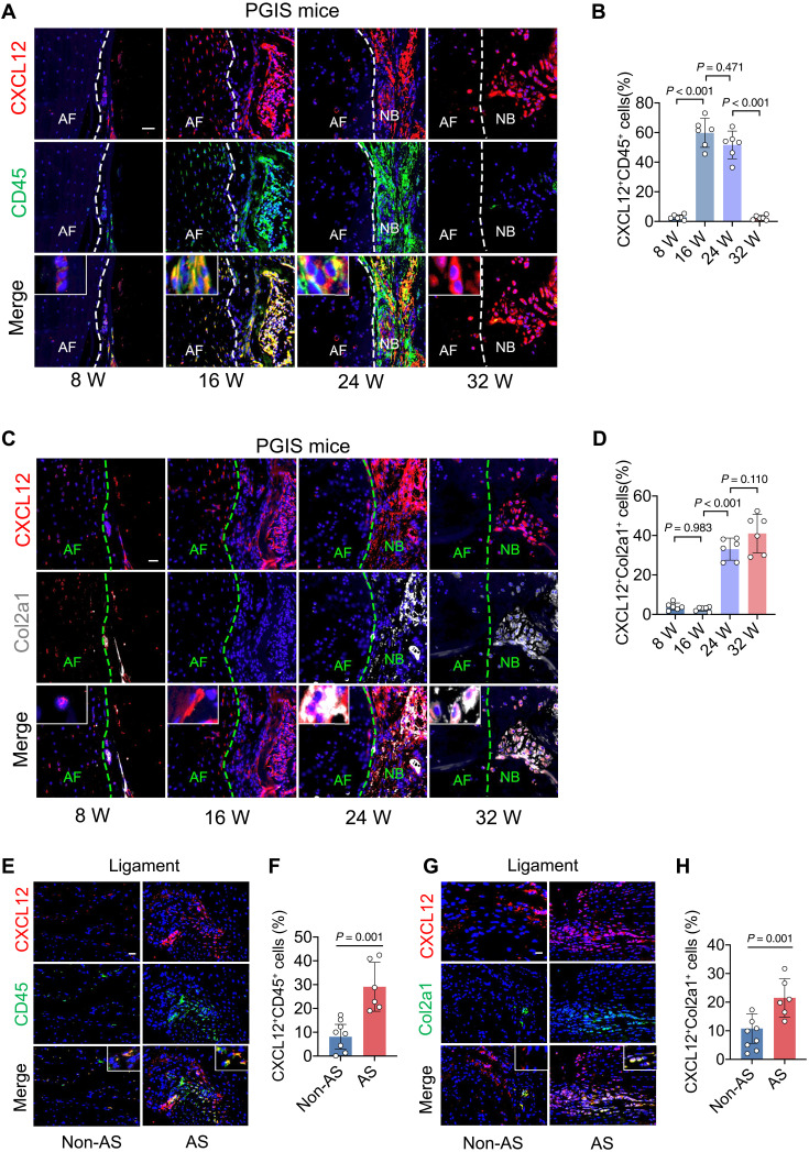
Fig. 5. CXCL12 is majorly produced by CD45+ cells in the inflammatory phase and Col2a1+ cells in the endochondral ossification phase.
(A and B) Immunohistochemical staining and quantitative analysis of CXCL12 and CD45 in PGIS mice at the age of 8, 16, 24, and 32 weeks. Scale bar, 20 μm. n = 6 per group. ANOVA (F3,20 = 121.10) with Tukey’s post hoc test was used. (C and D) Immunohistochemical staining and quantitative analysis of CXCL12 and Col2a1 in PGIS mice at the age of 8, 16, 24, and 32 weeks. Scale bar, 20 μm. n = 6 per group. ANOVA (F3,20 = 71.24) with Tukey’s post hoc test was used. (E and F) Immunohistochemical staining and quantitative analysis of CXCL12 and CD45 in spinal ligament tissues of patients with AS and non-AS patients. Scale bar, 20 μm. n = 6 tissues from patients with AS versus n = 8 tissues from non-AS patients. Student’s t test with Shapiro-Wilk test was used. (G and H) Immunohistochemical staining and quantitative analysis of CXCL12 and Col2a1 in spinal ligament tissues of patients AS and non-AS patients. Scale bar, 20 μm. n = 6 tissues from patients with AS versus n = 8 tissues from non-AS patients. Student’s t test with Shapiro-Wilk test was used. Data are shown as means ± SEM.

