. 2022 Mar 29;11:e75545. doi: 10.7554/eLife.75545

Figure 6. Glypican-1 (GPC1) and fibroblast growth factor 2 (FGF2) form a strong pair of interaction partners.

Recombinant constructs encoding soluble ectodomains of GPC1 (panel A), GPC1 ΔHS (panel B; a mutant form to which heparan sulfate chains cannot be added), GPC5 (panel C), GPC6 (panel D), and SDC4 (panel E; a member of the syndecan family of heparan sulfate proteoglycans) were expressed and purified from HEK293 cells (see Figure 6—figure supplement 1). Using biolayer interferometry, interactions studies with temporal resolution visualizing both association and dissociation kinetics were conducted with purified FGF2 (Figure 6—figure supplement 1) at the concentrations indicated. The data shown are representative for two independent experiments. Experimental details are given in the Materials and methods section.

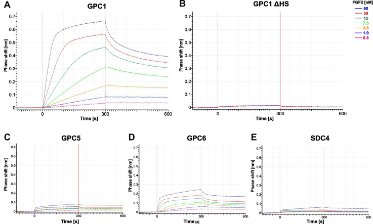
Figure 6.

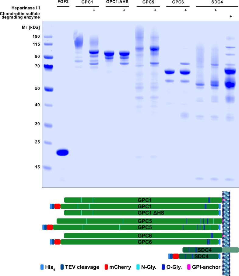
Figure 6—figure supplement 1. Expression and purification of soluble recombinant forms of fibroblast growth factor 2 (FGF2) and the various heparan sulfate proteoglycans.

Figure 6—figure supplement 1.

FGF2 was expressed and purified from Escherichia coli cells. All heparan sulfate proteoglycans indicated were expressed and purified from HEK293 cells. For details, see Materials and methods. Upper panel: SDS-PAGE analysis of all purified proteins (Coomassie staining). Lower panel: Schematic description of constructs used in this study in comparison to the endogenous forms.
Figure 6—figure supplement 1—source data 1. Unprocessed and uncropped image file of a SDS-PAGE analysis of the recombinant forms of GPC family members and fibroblast growth factor 2 (FGF2) used in the BLI experiments shown in Fig.
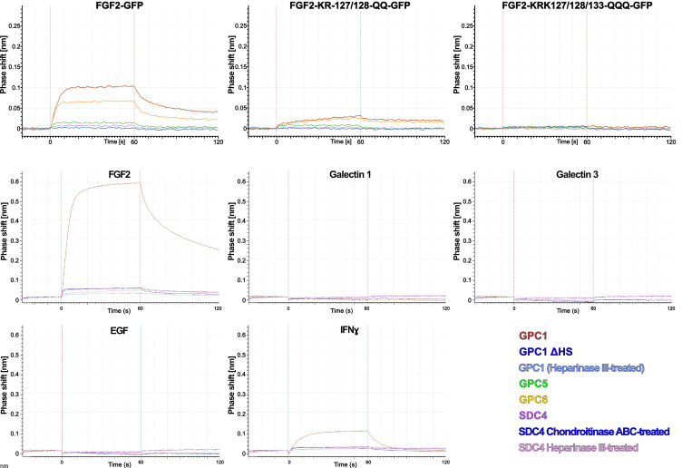
Figure 6—figure supplement 2. Interaction studies between different kinds of heparan sulfate proteoglycans and growth factors or cytokines using biolayer interferometry.

Figure 6—figure supplement 2.

Heparan sulfate proteoglycans were loaded onto BLI sensors as indicated by the color code. In some cases, they were treated with heparinase III or chondroitinase ABC as indicated. The binding partners were used in solution at a concentration of 60 nM. The data shown are representative for two independent experiments. For details, see Materials and methods.