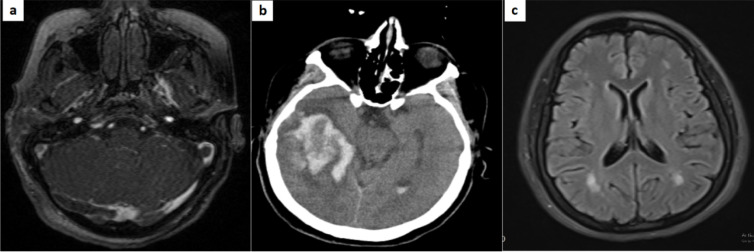
Figure2.
(a, b) Large hyper dense heterogeneous lesion in right temporal lobe with peripheral edema, more evaluated with brain magnetic resonance imaging/venography (MRI/MRV), which showed abnormal signal in right sigmoid sinus compatible with cerebral venous thrombosis; (c) T2 Flair images in a 39-year-old female with COVID-19 shows some hyper intense predominantly subcortical and deep white matter lesions without periventricular and corpus callosum involvement suggestive of acute disseminated encephalomyelitis (ADEM).

