
Extended Data Fig. 7. RC-COF-1 crystallite with photo-deposited Pt co-catalyst nanoparticles.
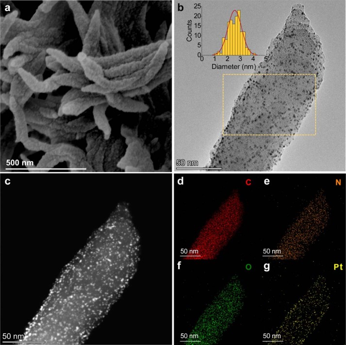
a–g, SEM (a), TEM (b), high angle annular dark field scanning transmission electron microscopy (HAADF-STEM) (c) images and elemental mapping (d–g) for RC-COF-1 crystallite decorated with photo-deposited Pt co-catalyst. The inset in b shows uniform distributions of Pt nanoparticles (2.5 ± 0.5 nm) in the selected area (yellow square). The uniform morphology of the reconstructed COF and the good Pt cocatalyst dispersion might also contribute to its enhanced activity.