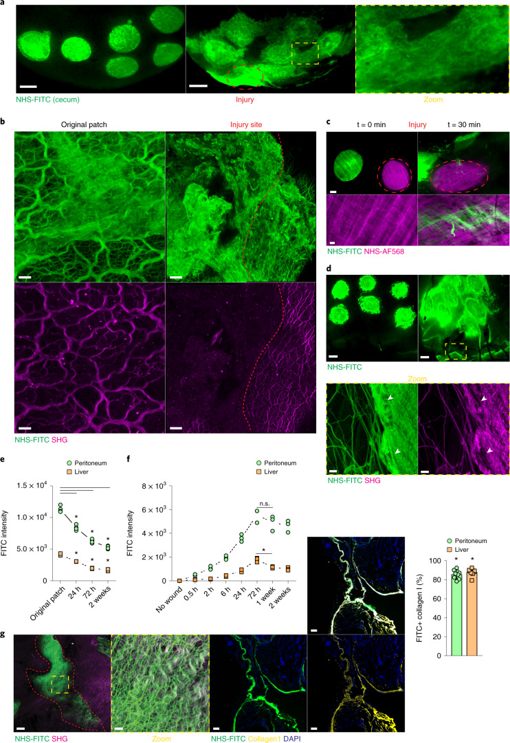
Fig. 1. Surface injury induces organ-wide matrix transfer.
a, Representative images from a liver surface marked with NHS-FITC at 24 hours p.i. Scale bar, 2,000 µm. b, Representative images of transferred matrix from the original patch and at a distal wound site. Wound area depicted in yellow. Scale bars: wound, 50 µm; original patch, 15 µm. c, Representative images of fate mapping of peritoneal surface ECM after brushing injury. Scale bars: overview, 1,000 µm; high magnification, 100 µm. d, Representative images showing that peritoneal surface ECM flows towards the laparotomy site. Scale bars: stereomicroscope, 2,000 µm; multiphoton, 15 µm. e, Fluorometric measurements of transferred matrix from original patch sites over time. n = 4 biological replicates (C57BL/6J wild-type (WT) mice). Data represent mean ± s.d. One-way ANOVA was used for the multiple comparisons testing, with Tukey’s test: *P < 0.05. f, Fluorometric measurements of transferred matrix into distal wound sites over time. n = 3 biological replicates (0.5 h, 2 h, 24 h, 2 weeks) and n = 4 (no wound, 6 h, 72 h, 1 week), (C57BL/6J WT mice). Data are mean ± s.d.. Two-tailed Mann–Whitney: *P < 0.05. n.s., P = 0.3429. g, Laparotomy closure in matrix fate mapping after 2 weeks. Scale bar: overview, 50 µm; high magnification, 15 µm. Percentage of FITC+ collagen I at the distal laparotomy site after 2 weeks. Scale bar, 50 µm. Data represent mean ± s.d. Two-tailed Mann–Whitney: *P < 0.05. Data in a–d and g were selected as representative of six biological replicates (C57BL/6J WT mice) and three independent experiments.

