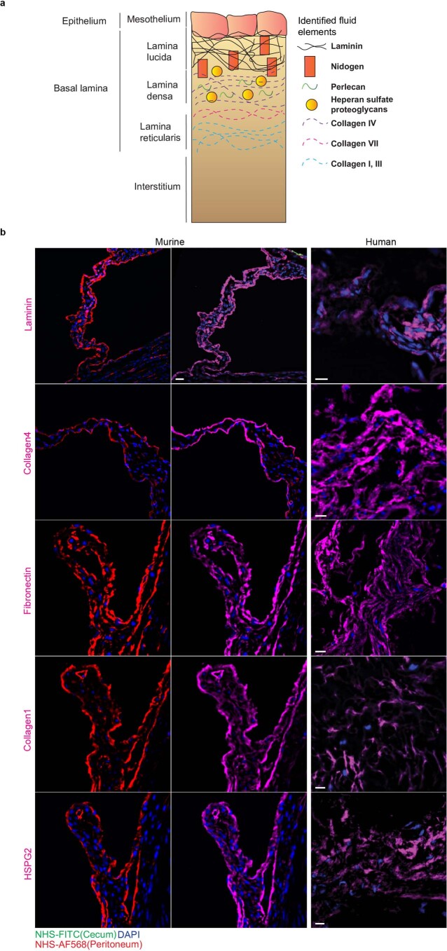
Extended Data Fig. 3. Fibrous postsurgical adhesions consist of transferred matrix elements.
a, Schematic Visualization of adventitial and serosal connective tissue layers identified transferred matrix. b, Representative immunofluorescence images of histological sections of three biological replicates of murine and human abdominal postsurgical adhesions. Murine peritonea were labeled with NHS-AF568 cecum was labeled with NHS-FITC. Mice were sacrificed 4 weeks after surgery. n = six C57BL/6 J WT mice and four independent experiments. Human adhesion tissue was collected from ten different patients with abdominal adhesions during adhesiolysis. Scale bar: 20 µm.

