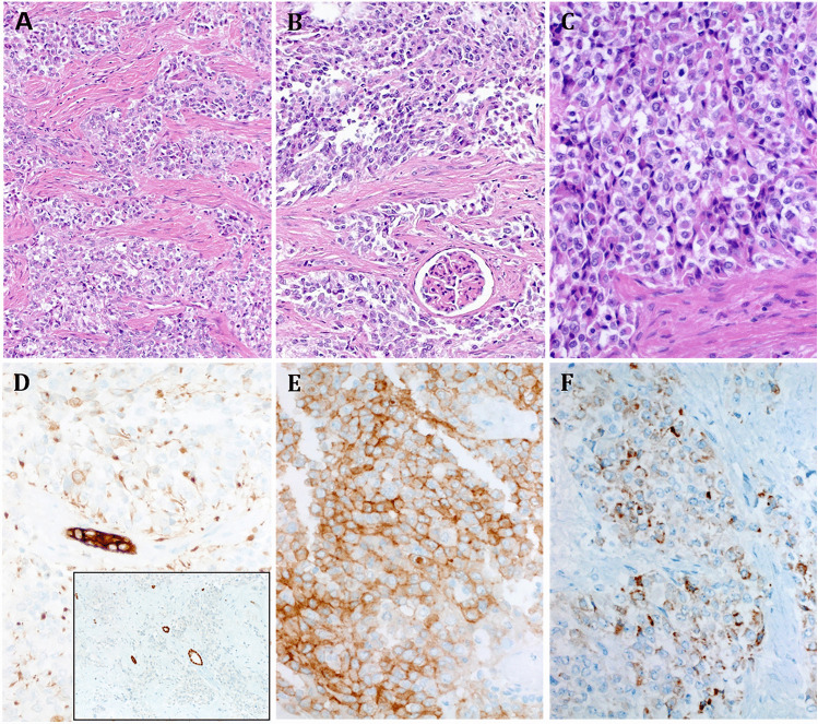
Fig. 1.
Representative histological images of Case 1 show epithelioid cells disposed into communicating irregular nests within fibrous stroma (A) with focal entrapment of the glomeruli (B). C At high power, the epithelioid morphology is seen; note the variably granular eosinophilic to clear cytoplasm. D AE1/AE3 reveals mainly paranuclear dot-like pattern (note the strong expression in entrapped tubules; main image). D, inset, PAX8 highlights entrapped tubules, but the tumor cells are negative. E Strong cytoplasmic ALK expression is seen. F MUC4 is variably positive

