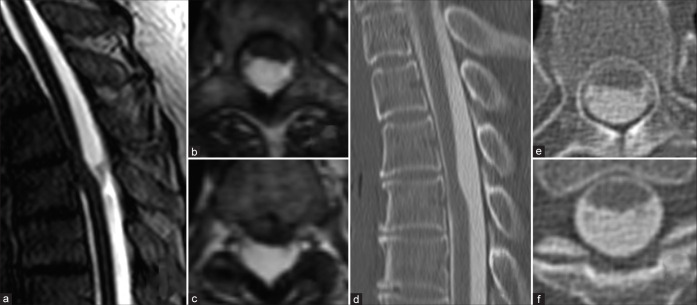
Figure 1:
MRI T2 CISS revealed anterior subarachnoid space of the spinal cord was not clear and relatively focal ventral shifting of the spinal cord. (a-c) Sagittal image of CT myelography showed ventral CSF space but coronal image of CT myelography showed interruption of ventral CSF space. (d-f). Abbreviation; CISS: Constructive interference in steady state, CT: Computed tomography, CSF: Cerebrospinal fluid.

