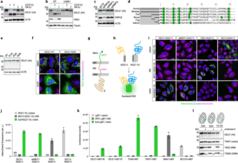
Fig. 2. DELE1 is a substrate of MPP that can be fully imported into the mitochondrial matrix.
a HeLa DELE1HA cells treated as indicated and analyzed for endogenous DELE1 protein by immunoblotting. OM, oligomycin. Blue, L-DELE1. Green, M-DELE1. Pink, S-DELE1. b 293 T DELE1HA WT and OMA1 knockout (ΔOMA1) cells treated as indicated and analyzed by immunoblotting. c HeLa DELE1HA cells exposed to sgRNAs targeting the indicated genes for six days and analyzed by immunoblotting. d Alignment of the first 50 amino acids of DELE1 from different species. Arginines highlighted (green). e Wild-type DELE1 or the indicated arginine to lysine mutants transiently expressed in HeLa cells. Cell lysates analyzed by immunoblotting. f Confocal microscopy of HeLa cells transfected with indicated DELE1 variants. Scale bars,10 μm. Nuclei (DAPI, blue), mitochondria (MitoTrackerRed, pink), DELE1 (HA, green). g Schematic illustrating DELE1 import into mitochondria and its processing by MPP and OMA1. IMM, inner mitochondrial membrane. IMS, intermembrane space. OMM, outer mitochondrial membrane. h Principle of split mNeonGreen2 (NG2) complementation by fusion of the small (NG2(11)) and the large (NG2(1-10)) component to different proteins (yellow, blue) sorted into the same subcellular compartment. i Localization of DELE1 in HeLa cells analyzed via split NG2 system by confocal microscopy. NG2(11) was C-terminally fused to DELE1, altMIEF1, SEC13 or the sorting signal of PSD1 and co-expressed with NG2(1-10) directed to different subcellular compartments by fusion with the sorting signals of MICU1 or Su9. Scale bars, 20 μm. Nuclei (DAPI, blue), mitochondria (MitoTrackerRed, pink), mNG2 (green). j NG2 fluorescence as in (i) measured by flow cytometry. Mean ± s.d. of n = 3 independent biological samples. (k) Localization of endogenous DELE1 assessed by split luciferase reporter system. HeLa cells expressing the indicated endogenous proteins tagged with the small luciferase fragment (HiBiT) were transfected with the large luciferase fragment (LgBiT), unmodified or fused to the sorting signals of OPA1 or Su9. Luminescence measured 24 h post-transfection. Mean ± s.d. of n = 3 independent biological samples. l Accessibility of proteins to proteinase K in mitochondria isolated from HeLa DELE1HA cells in isotonic buffer (isoto.), upon osmotic swelling (hypotonic buffer, hypo.) or lysis of mitochondria (TX-100).

