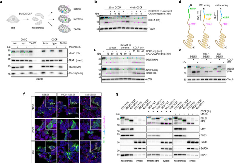
Fig. 3. Release of DELE1 into the cytosol is coupled to sorting and maturation.
a Mitochondria isolated from 293T DELE1HA OMA1 knockout (ΔOMA1) cells treated with DMSO or CCCP for 4 h were processed as in Fig. 2l. b, c HeLa DELE1HA cells (b) and HeLa cells stably expressing DELE1-HA (c) were pretreated with CHX to deplete newly synthesized DELE1 protein, followed by co-treatment of CHX with CCCP for the indicated times and analysis of DELE1 protein by immunoblotting. CCCP only and simultaneous CHX + CCCP treatment without CHX pretreatment serve as controls. d Schematic comparing wild-type DELE1 and chimeric DELE1 proteins carrying a heterologous MTS and their associated protease cleavage sites, used in the subsequent panels. e 293T DELE1 knockout cells stably expressing the indicated DELE1 variants were treated with CCCP or OM for 6 h. Processing of the DELE1 proteins was analyzed by immunoblotting. f 293T DELE1 knockout cells were transiently transfected with the specified DELE1 chimeras, treated for 4 h as indicated and analyzed by confocal microscopy. Scale bars, 20 μm. Nuclei (DAPI, blue), mitochondria (MitoTrackerRed, pink), DELE1 (HA, green). g Subcellular fractionation reveals the localization of different DELE1 species to mitochondria or cytosol upon CCCP or OM treatment in 293T DELE1 knockout cells transiently transfected with the indicated variants.

