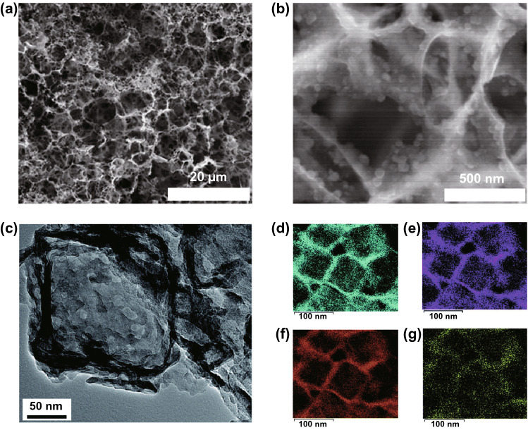
Fig. 13.
Scanning electron micrographs of a 3D porous carbon scaffold onto which sulphur nanoparticles are deposited, at a low magnification and b high magnification [180]. Creative Commons License (CC BY 4.0). c Transmission electron micrograph of a polysulphide-adsorbing TiO2-MnO nanobox cathode infiltrated with sulphur, and associated energy-dispersive X-ray spectroscopy maps for d Ti, e Mn, f O and g S [181].
Copyright 2021, Royal Society of Chemistry

