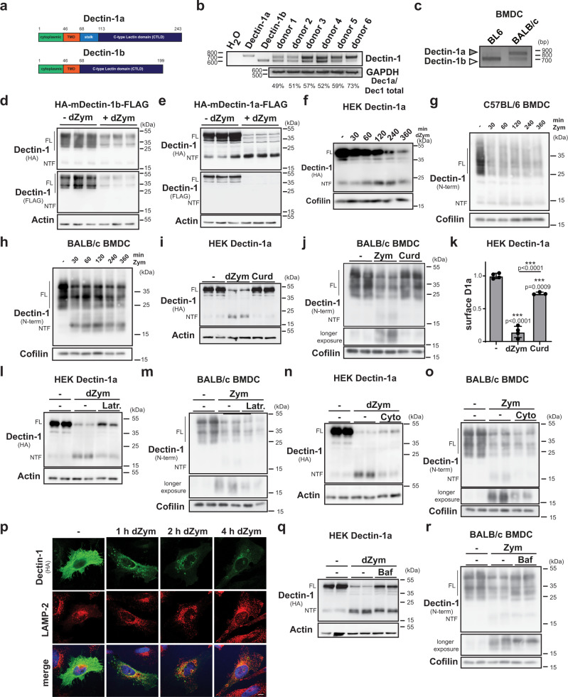
Fig. 1. Differential degradation of Dectin-1 isoforms.
a Scheme of murine Dectin-1 proteins. b Expression of Dectin-1 isoforms in human PBMCs was analysed by RT-PCR. N = 1, n = 6. c cDNA generated from RNA isolated from BMDCs of either C57BL/6 (BL6) or BALB/c mice was subjected to RT-PCR using Dectin-1-specific primers. N = 3, n = 3. d HEK cells stably overexpressing HA-mDectin-1b-FLAG were treated for 6 h with 50 µg/ml depleted Zymosan (dZym) or left untreated and subjected to western blot analysis. Full-length (FL) Dectin-1 and the corresponding N-terminal fragment (NTF) are highlighted throughout the Figure. N = 2, n = 6. e The experiment described in d) was repeated with HEK cells stably transfected with HA-mDectin-1a-FLAG. N = 2, n = 6. f Dectin-1a expressing HEK cells were treated for 0, 30, 60, 120, 240 or 360 min with 50 µg/ml dZym and analysed by western blotting. N = 3, n = 3 BMDC from C57BL/6 (g) or BALB/c (h) mice were stimulated for the indicated time points with 100 µg/ml Zymosan (Zym) and analysed for processing of Dectin-1 by western blotting. N = 1, n = 3 in both cases. i Dectin-1a expressing HEK cells were treated for 6 h with 50 µg/ml dZym or 200 µg/ml Curdlan (Curd) and subjected to western blot analysis. N = 3, n = 6. j The experiment in i was repeated with BALB/c BMDCs treated with 100 µg/ml Zym instead of dZym. N = 3, n = 6. k Surface expression of Dectin-1a was analysed in the setup described in i by flow cytometry. Bars depict Mean ± SD N = 2, n = 3 (Curd), n = 4 (dZym). One-way ANOVA with Tukey’s post hoc testing. Stably Dectin-1a expressing HEK cells (l N = 3, n = 6) or BALB/c BMDCs (m N = 3, n = 6) were treated for 30 min with 5 µM Latrunculin A (Latr.) prior to stimulation with 50 µg/ml dZym or 100 µg/ml Zym for 6 h and detection of Dectin-1 by western blotting. The experiment was repeated in the same cells but with pre-treatment with 1 µM Cytochalasin D (Cyto) (n, o). N = 3, n = 6 in both cases. p HeLa cells transfected with HA-mDectin-1a-FLAG were treated with 50 µg/ml dZym, fixed and subjected to immunofluorescence analysis. Scale bar, 10 µm. N = 3, n = 3 HEK cells overexpressing HA-mDectin-1a-FLAG (q N = 3, n = 6) or BALB/c BMDC (r, N = 3, n = 6) were pretreated for 30 min with 100 nM Bafilomycin A1 (Baf) and stimulated for 30 min with 50 µg/ml dZym or 100 µg/ml Zym prior to western blot analysis. ***p ≤ 0.001.

