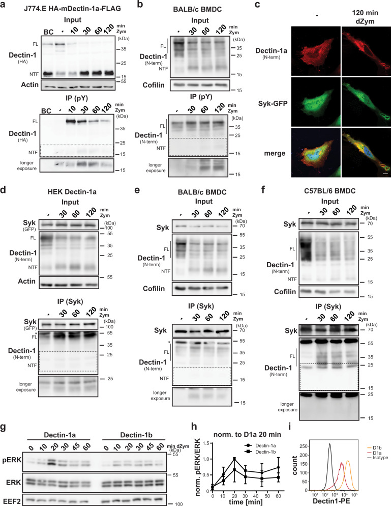
Fig. 2. The Dectin-1a NTF persists in a signalling-competent state.
a J774.E cells stably expressing HA-mDectin-1a-FLAG were stimulated for the indicated time points using 100 µg/ml Zymosan (Zym). Lysates from cells treated for 20 min were employed as bead control (BC) where no precipitating antibody was added. After lysis, tyrosine-phosphorylated (pY) proteins were immune-precipitated (IP) using a phospho-Tyr antibody and protein G agarose beads. Lysates (input) and pull-down samples were analysed by western blotting. Full-length (FL) Dectin-1 and the corresponding N-terminal fragment (NTF) are highlighted throughout the Figure. N = 3, n = 3. b BMDC from BALB/c mice were subjected to the same experimental procedure described in a. N = 3, n = 3. c Co-localisation of Syk-GFP and HA-mDectin-1a-FLAG was validated in transiently transfected HeLa cells. Cells were treated for the indicated time points with 50 µg/ml depleted Zymosan (dZym) and subjected to immunofluorescence analysis using a Dectin-1-specific antibody. Scale bar, 10 µm. N = 3, n = 3. d Association of the Dectin-1a NTF with Syk was analysed in HEK cells stably expressing HA-mDectin-1a-FLAG transiently transfected with Syk-GFP. After lysis, Syk was precipitated with a specific antibody and protein G agarose beads and lysates as well as IP samples were subjected to western blot analysis employing the indicated antibodies. N = 3, n = 3. e Interaction of Syk and the Dectin-1a NTF was validated in BMDC of BALB/c wild type mice treated for 0, 30, 60 or 120 min with 100 µg/ml Zymosan. Syk was precipitated from lysates using a specific antibody and lysates and IP samples were analysed by western blotting using the indicated antibodies. N = 3, n = 3. f The experiment described in e was performed using BMDC isolated from C57BL/6 mice. N = 2, n = 4. g HEK cells stably overexpressing HA-mDectin-1a-FLAG or HA-mDectin-1b-FLAG were serum-starved for 1 h and subsequently stimulated for the indicated time points with 50 µg/ml dZym. Phosphorylation of ERK1/2 was finally evaluated by western blotting. N = 6; n = 6. h Quantification of g. N = 6; n = 6. Bars indicate Mean±SD. i Dectin-1 surface expression of the cell lines used in h was analysed by flow cytometry using a Dectin-1-specific antibody. N = 6, n = 6.

