
Figure 2.
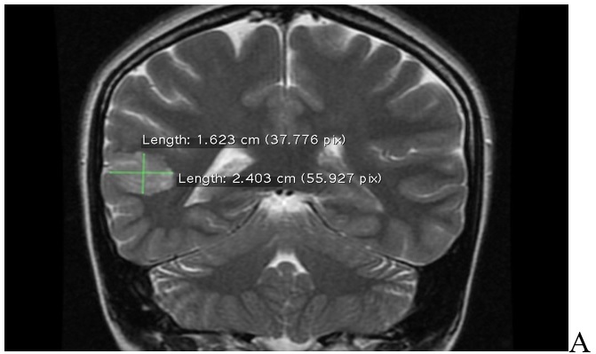
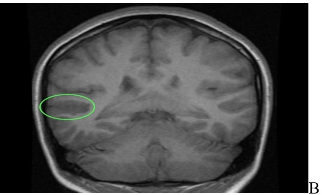

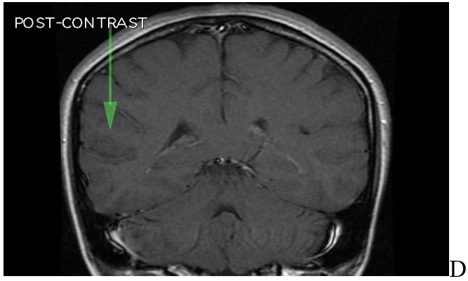
MRI of brain. A: T2 sequence coronal plane results: abnormal area of cortical dysplasia is measured about 24x16mm, consistent with Blumcke Type I (Taylor’s type) cortical dysplasia, “without transmantle extension” towards ventricle. B: Sequence T1 axial plane without contrast results: cortical thickening up to 5mm on either side of posterior aspect of right superior temporal sulcus (involving the posterior aspects of both superior temporal gyrus and middle temporal gyrus). This abnormal area of cortical dysplasia is measured about 24x16mm. C: T1 sequence sagital plane post contrast, no pathologic contrast enhancement is identified. D: T1 sequence coronal plane post contrast, no pathologic contrast enhancement is identified