
Fig. 1.
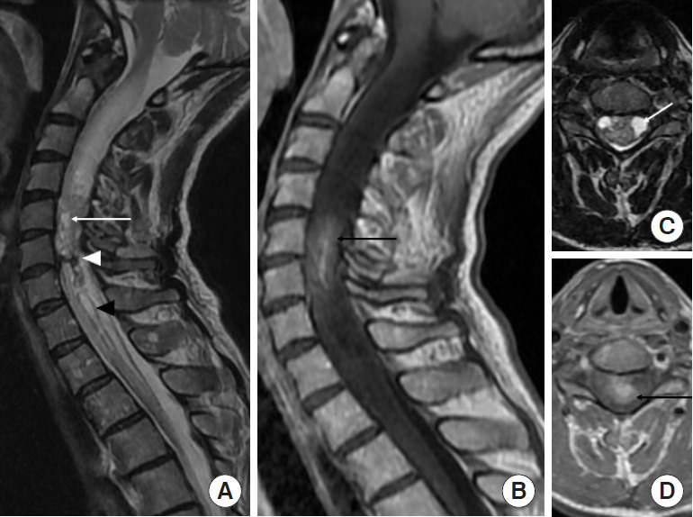
Ependymoma (World Health Organization grade II) in a 57-year-old woman. (A, C) Sagittal and axial T2-weighted images reveal a heterogeneous expansile intramedullary mass in the cervical spine, with intratumoral cysts (white arrow) and hemosiderin cap sign (white arrowhead) at the inferior margins of the mass. Surrounding edema is present associated with syringohydromyelia (black arrowhead). (C) Axial T2-weighted image confirms the central location in the spinal cord. (B, D) Sagittal and axial contrast-enhanced T1-weighted images show intratumoral enhancement (black arrow) at C5–6 levels.