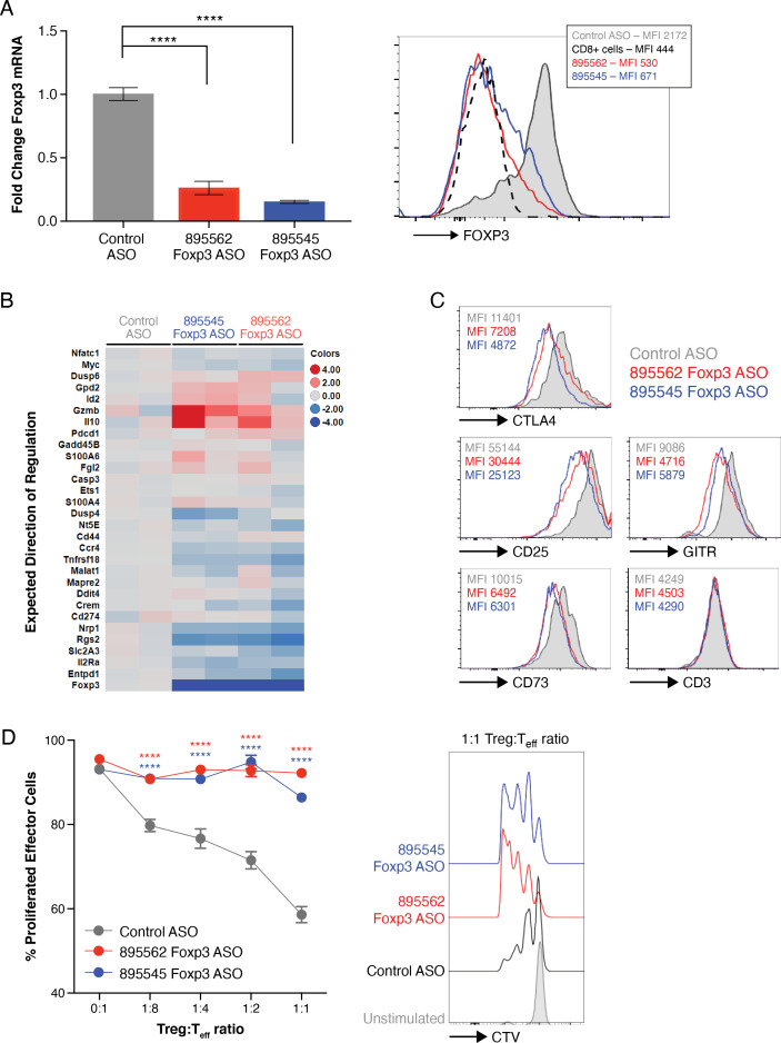
Figure 2.
Antisense-mediated knockdown of mouse FOXP3 promotes loss of Treg suppressive phenotypic markers and function. (A) Primary Tregs were isolated from mouse spleen and cultured with ASOs (5 µM) in triplicates for 7 days. Bar chart shows relative FOXP3 mRNA expression measured by RT-qPCR, histogram shows FOXP3 protein expression measured by flow cytometry. (B) Inducible Tregs (iTregs) were differentiated and cultured in the presence of ASO for a total of 7 days. Heatmap shows expression of FOXP3-dependent mRNA genes as measured by Fluidigm. (C) Histograms show abundance of indicated proteins in primary Tregs. (D) iTregs were differentiated and cultured in the presence of ASOs and evaluated for their ability to inhibit proliferation of effector cells in an in vitro suppression assay in duplicates. Histograms show representative CellTrace Violet (CTV) staining on effector cells cultured at a 1:1 Treg:Teffector ratio. Data in figure represent ≥2 independent experiments. *, P≤0.05; **, p≤0.01; ***, p≤0.001; ****, p≤0.0001 by one-way analysis of variance (ANOVA) with Dunnett’s post-test for (A) and two-way ANOVA with Dunnett’s post-test for (D). Differences are calculated relative to control ASO. ASOs, antisense oligonucleotides; mRNA, messenger RNA; Tregs, regulatory T cells.

