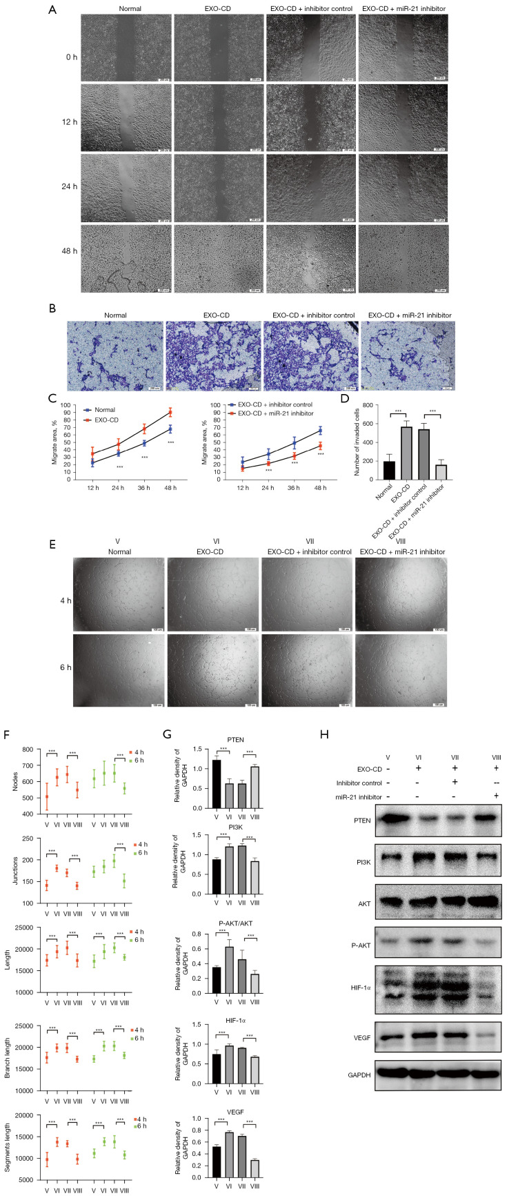
Figure 3.
In vitro, miR-21 inhibitors can effectively antagonize the effect of exosomes on promoting vascular endothelial migration and tubule formation in patients with CD. (A,C) Wound healing assay. Observational method: differential interference contrast microscope. (B,D) Transwell migration assay. Cells are stained with crystal violet. (E,F) Tube formation assay. Observational method: phase contrast microscope. (G,H) Protein expression of the PTEN/PI3K/AKT/HIF-1a/VEGF axis analyzed by western blot. Densitometry was used to quantify proteins expression relative to GAPDH. Roman numerals represent different processing groups. V, Normal. VI, Exo-CD, plasma-derived exosomes from patients with CD. VII, Exo-CD + miRNA inhibitor controls. VIII, Exo-CD + miR-21 inhibitors. ***P<0.001. The results were repeated 3 times. CD, Crohn’s disease; PTEN, phosphatase and tensin homolog; PI3K, phosphoinositide 3-kinase; AKT, AKT serine/threonine kinase; HIF-1α, hypoxia-inducible factor 1-alpha; VEGF, vascular endothelial growth factor; GAPDH, glyceraldehyde 3-phosphate dehydrogenase; miRNA, microRNA; miR-21, microRNA 21.

