FIGURE 7.
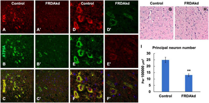
The number of frataxin- and ATP5A-positive-cerebellar dentate nucleus principal neurons is decreased in FRDAkd mice at 18 weeks of dox treatment. Confocal images of frataxin (FXN) (red) and mitochondrial marker ATP5A (green) fluorescence, and merged images with DAPI-stained nuclei (blue) in the cerebellum of FRDAkd (A′–F′) and control (A–F) mice. Hematoxylin-Eosin staining of cerebellar principal neurons FRDAkd (H) and control mice (G). (I) Quantification of ATP5A-positive principal neurons in FRDAkd and control mice at 18 weeks of dox treatment (Stats: n = 3 mice per group, 3 sections per mouse, mean ± SEM, student’s t-test, **P < 0.01).

