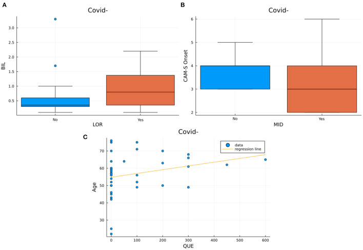
Figure 4.
Visualization of the significant results from the correlations in the COV–/DEL+ group. Box plots represent the bilirubin levels distributions in subjects assuming LOR vs. subjects not assuming LOR (Figure 3A) and the distribution of different CAM-S scores at the onset of delirium in subjects assuming MID and in subjects not assuming MID (Figure 3B). The scatter plot with regression line (Figure 2D) represent age vs. maximum dose of quetiapine used.

