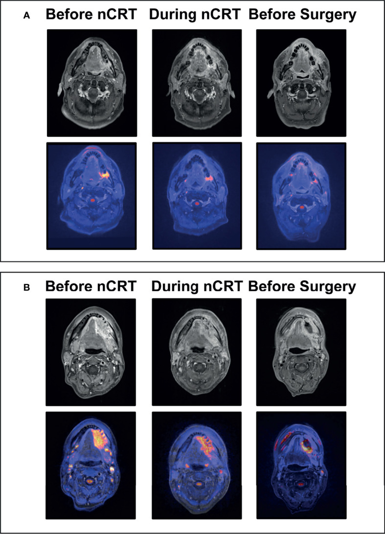
Figure 2.

Exemplary MRI Images of Clinical Responses to Neoadjuvant Chemoradiotherapy. (A) Exemplary images of a 55-year old patient with left-sided squamous cell carcinoma of the oral cavity before and during chemoradiotherapy (day 15), and prior to surgery; The top row shows representative axial gadolineum-enhanced T1-weighted images with continuous decrease in size and contrast enhancement resulting in complete clinical response prior to surgery of the primary tumor at the left retromolar region; The bottom row shows corresponding fused diffusion-weighted - gadolineum-enhanced T1-weighted images with decreasing diffusion restriction of the tumor region resulting in complete clinical response prior to surgery. (B) Exemplary images of a 49-year old patient with left-sided squamous cell carcinoma of the oral cavity before and during chemoradiotherapy (day 15), and prior to surgery; The top row shows representative axial gadolineum-enhanced T1-weighted images with continuous decrease in size and contrast enhancement. Markable residual tumor with contrast enhancement at the left mandibular region prior to surgery; The bottom row shows corresponding fused diffusion-weighted - gadolineum-enhanced T1-weighted images with decreasing but residual diffusion restriction of the tumor region; nCRT, Neoadjuvant chemoradiotherapy.
