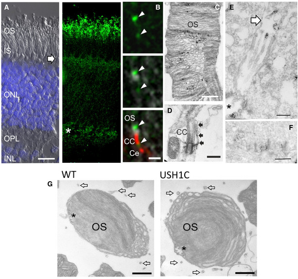
Representative staining of longitudinal sections from porcine WT retina (2 pigs, 1 retina each, 4 TR).
-
A
Harmonin (green) is present in the layer of PRC outer segment (OS), the outer limiting membrane (arrow) and in the outer plexiform layer (OPL, asterisk), where PRC synapses are located. Left image: DIC image super‐exposed with DAPI for nuclei in outer nuclear layer (ONL) and inner nuclear layer (INL). Right image: Immunohistological staining with anti‐harmonin antibody. IS, PRC inner segment. Scale bar 10 µm.
-
B
Immunohistological co‐localization of harmonin (green) and the connecting cilia (CC)/centriole (Ce) marker centrin (red) at the OS base (arrowheads, scale bar 1 µm).
-
C, D
(C) Immunoelectron microscopy detects harmonin at PRC OS discs (scale bar 500 nm) as well as (D) at the base of connecting cilia (CC, scale bar 500 nm) and in calyceal processes (arrows).
-
E, F
(E) In Müller glia cells, harmonin is localized at microvilli tips (arrow, scale bar 500 nm), at the cell adhesion region (asterisk) as well as (F) in cone synaptic pedicles (scale bar 500 nm).
-
G
Horizontal cross sections through cone OS reveal persistence of calyceal processes in USH1C pig (arrows, scale bar 400 nm). Asterisks indicate axoneme projections into the OS.

