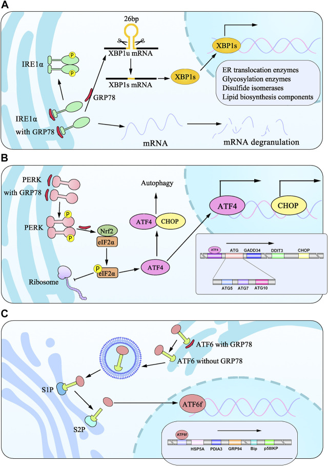
FIGURE 1.
Model diagram of signaling pathways from three ER transmembrane stress sensors during UPR induced by ER stress. (A) IRE1α isolates from BiP undergo dimerization and phosphorylation to splice 26bp from XBP1 into XBP1s, which translocates to the nucleus and induces the transcription of target genes. (B) PERK signaling is initiated through PERK dimerization and autophosphorylation of the cytosolic PERK kinase domain. eIF2α phosphorylation is elicited to attenuate global protein synthesis. eIF2α promotes translation of ATF4, which translocates to the nucleus to regulate the expression of target genes and cooperates with CHOP to participate in ER stress-induced apoptosis pathways. (C) ATF6 dislocates from BiP translocates to Golgi apparatus, which is cleaved by proteases at S1P and S2P sites into ATF6f and promotes the transcription of genes.

