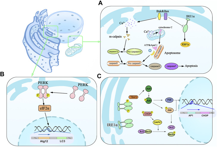
FIGURE 3.
Schematic diagram of apoptosis associated with the mitochondrial pathway and autophagy induced by ER stress. (A) Effector proteins of Bax and Bak in apoptosis undergo conformational changes in active forms and oligomerize in the ER membrane leading to the efflux of Ca2+ from the ER to the cytoplasm and activating m-calpain to cleave and activate procaspase-12, which in turn activates caspase-3 and caspase-7. BAX and BAK are associated with the decreased expression of IRE1 and XBP1 and promote apoptosis. Ca2+ can be taken up by mitochondria leading to depolarization of the mitochondrial inner membrane and cytochrome c release, which binds Apaf-1, procaspase-9, and ATP into apoptosome and activates caspase-9 and caspase-3, then resulting in cell death. (B) Dimmerized PERK phosphorylates the translation initiation factor eIF2α and mediates the translation of Atg12 and LC3. (C) Activated IRE1 forms P-IRE1α/TRAF2 complex and initiates Ask1 as a stress-responsive in the JNK and p38 pathways. Phosphorylated JNK along with activated P38 enters the nucleus to promote translation of AP1 and CHOP.

