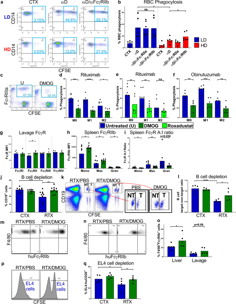
Fig. 7.
The impact of hypoxia-driven FcγRIIb upregulation on mAb mediated target cell depletion. a, Flow cytometry plots showing levels of uptake of CSFE+ red blood cells (RBCs) by LD and HD monocytes. RBCs sourced from Rhesus D+ individuals were opsonised with control cetuximab (CTX) or anti-Rhesus D antigen specific mAb (αD). RBCs used as targets for LD and HD pre-cultured monocytes pre-treated with or without anti-FcγRIIb (αFcγRIIb) blocking mAb. b, RBC phagocytosis quantified for 6 donors. c, Flow cytometry plots showing Rituximab mediated uptake of CLL cells by FcγRIIIa+ M1 macrophages generated with or without DMOG. d, CLL cells opsonised with Rituximab and cultured with M0, M1 or M2 MDMs generated in the absence or presence of DMOG or e, Roxadustat and the percentage of phagocytic MDMs were determined by flow cytometry (n = 6–8 per group). f, Phagocytosis of CLL cells mediated by Obinutuzumab (n = 6 per group). g, FcγR expression on F4/80+ macrophages in the peritoneal lavage of WT C57BL/6 mice treated with DMOG or PBS control. i.p., determined using flow cytometry (n = 6 per group). h, FcγRIIb expression levels and i, FcγR A:I ratio were determined by flow cytometry in splenic monocytes (Mono), macrophages (Mac) and granulocytes (Gran) of DMOG or PBS treated hFcγRIIb/mFcγRIIKO mice (n = 8 per group). j, hFcγRIIb/mFcγRIIKO/hCD20 mice were treated with DMOG or vehicle PBS control i.p. for 72 h prior to receiving Rituximab (RTX) or CTX isotype control. %CD19+ cells in the peripheral blood of each mouse were determined using flow cytometry (n = 8–10 per group). k, hFcγRIIb/mFcγRIIKO mice were treated with DMOG or PBS i.p. for 72 h prior to receiving CFSE labelled target splenocytes from hCD20/mFcγRIIKO mice and non-target splenocytes from WT C57BL/6 mice, i.v. These mice were treated with DMOG or PBS i.p. prior to receiving RTX or CTX 24 h later. Flow cytometry plots are shown for the depletion of target and non-target splenocytes, and l, data is presented as CD19+ cell target:non-target ratio (n = 5 per group). m, Flow cytometry plots showing hFcγRIIb expression on liver and n, peritoneal lavage F4/80+ macrophages 72 h post-treatment with DMOG or PBS control, i.p., in hFcγRIIb/mFcγRIIKO mice, and o, quantified for 5 mice per group. p, hFcγRIIb/mFcγRIIKO mice were treated with DMOG or PBS i.p. for 60 h prior to receiving CFSE labelled EL4-huCD20+ cells following treatment with RTX or CTX, and DMOG or PBS. Histograms showing depletion of target EL4-huCD20+ cells in the peritoneum by RTX in the absence and presence of DMOG and q, EL4-huCD20+ cell depletion in the peritoneal lavage quantified using flow cytometry. Bars represent group means. Statistical significance was assessed using an unpaired two-tailed t-test, a paired two-tailed Wilcoxon test, or a one-way ANOVA for the in vivo cell depletion experiments (*p < 0.05, **p < 0.01, ***p < 0.001, ****p < 0.0001 and ns = non- significant). Also see Additional file 7: Fig. S7

