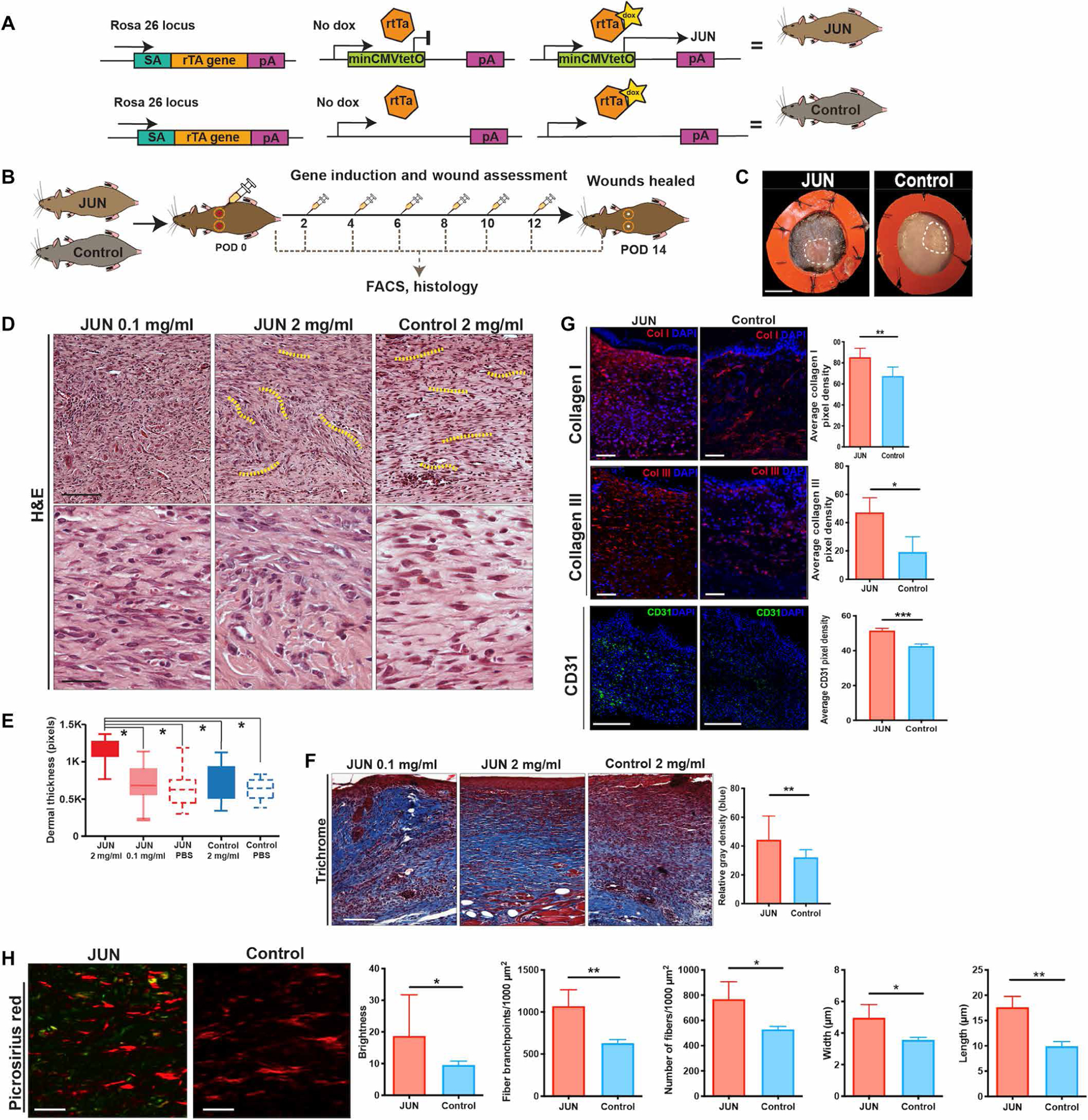
Fig. 1. Jun overexpression induces scar formation.

(A) Targeting construct of JUN doxycycline (dox)–inducible mice. In JUN mice, the construct at the Rosa 26 locus (rTtA) is coupled with the tetracycline operator minimal promoter (tetO) and leads to Jun overexpression in the presence of dox (top right) but no change to Jun expression without dox (top middle). In the Control mice, Jun is physiologically expressed due to the lack of the rtTA with (bottom right) and without dox (bottom middle). (B) Schematic showing experimental approach: Six-millimeter stented excisional dorsal wounds were created in JUN and Control mice. Dox (20 μl of 2 mg/ml) was administered on the day of surgery and on alternate postoperative days (POD) until complete wound closure (POD 14). Wounds were harvested for fluorescence-activated cell sorting (FACS) and histology (n = 18 mice per group per time point). (C) Representative gross photographs of healed (POD 14) wounds from JUN and Control mice receiving dox (2 mg/ml). White dotted line, healed scar. Scale bar, 0.25 cm. (D) Representative hematoxylin and eosin (H&E)–stained wounds of JUN and Control mice. Scale bars, 75 μm (top) and 25 μm (bottom). Yellow dotted lines show the whorl pattern of hypertrophic scars (HTSs). (E) Comparison of dermal thickness in wounds of JUN and Control mice at POD 14. (F) Representative Masson’s trichrome–stained wounds of JUN and Control mice at POD 14. Comparison of total collagen content (defined as relative mean gray density) from Masson trichrome staining. Scale bar, 100 μm. (G) Immunofluorescently labeled collagen type I (COL1) (red), COL3 (red), and CD31 (green) in JUN and Control mice on POD 14. Scale bars, 100 μm. DAPI, 4′,6-diamidino-2-phenylindole. (H) Picrosirius red–stained wounds of JUN and Control mice on POD 14. Scale bars, 25 μm. Computational quantification of collagen fiber networks evaluating length, branching, brightness, width, and number of fibers in JUN mice. All data are presented as means ± SEM. n = 3 independent experiments. *P < 0.05, **P < 0.01, and ***P < 0.001.
