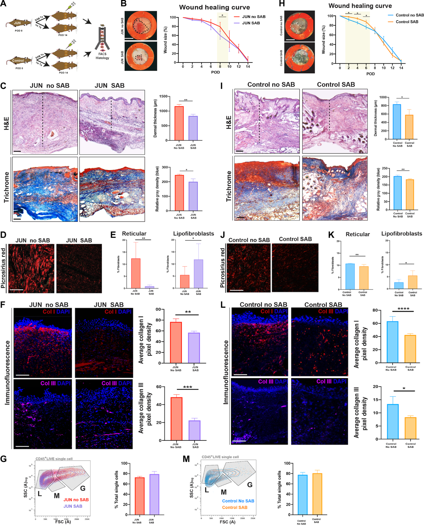
Fig. 5. CD36 antagonism reverses hypertrophic dermal scarring.

(A) Schematic of the experimental approach: Six-millimeter stented excisional dorsal wounds were created in JUN and Control mice. Dox (20 μl of 2 mg/ml) and SAB (100 μM) were administered on the day of surgery and on alternate PODs until complete wound closure (POD 14). Wounds were harvested for FACS and histology (n = 18 mice). Representative gross photographs of healed (POD 14) wounds of JUN (B) and Control (H) mice. Scale bars, 0.25 cm. Representative H&E- and Masson trichrome–stained wounds of JUN (C) and Control (I) mice. Scale bars, 150 μm. Picrosirius red–stained wounds of JUN (D) and Control (J) mice on POD 14. Scale bars, 25 μm. Sections are not adjacent slides but are from the same experimental group. Immunofluorescently labeled Col1 (red) and Col3 (purple) in JUN (F) and Control (L) mice on POD 14. Scale bars, 100 μm. Bar graph showing the numbers of lipofibroblasts and reticular fibroblasts in JUN (E) and Control (K) mice at POD 14. Bar graph showing the total percent of immune cells in JUN (G) and Control (M) mice at POD 14. All data are presented as means ± SEM. n = 3 independent experiments. *P < 0.05, **P < 0.01, ***P < 0.001, and ****P < 0.0001.
